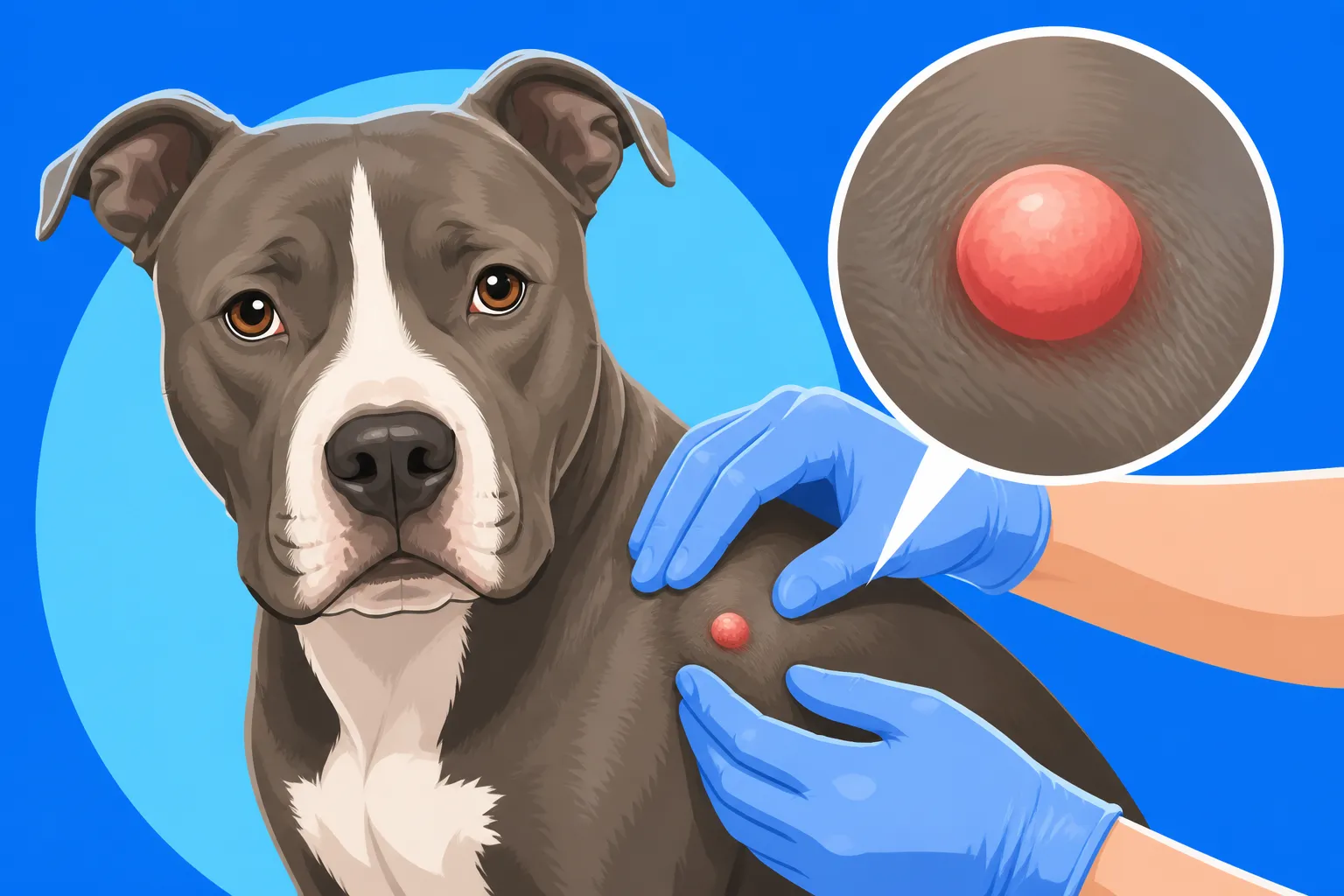
Feeling for a Mast Cell Tumor on a Pit Bull
Finding a new lump or bump on your beloved Pit Bull can send a wave of panic through any dog parent. It's a gut-wrenching moment, immediately making you wonder if it's something serious like a mast cell tumor or just a harmless fatty deposit. This guide is here to walk you through how to systematically check your Pit Bull for suspicious lumps and understand what characteristics might indicate a mast cell tumor, empowering you to take informed action for your dog's health.
Understanding Mast Cell Tumors in Pit Bulls
When we talk about bumps and lumps on our dogs, mast cell tumors are often among the most concerning for good reason. They can be tricky, mimicking all sorts of benign growths, which is why being vigilant and knowing what you're looking for is so crucial, especially with a breed like the Pit Bull.
What are Mast Cell Tumors?
Mast cells are a normal part of your dog's immune system, playing a role in allergic reactions and inflammation. They're found throughout the body, particularly in the skin, respiratory tract, and gastrointestinal tract. A mast cell tumor, then, is essentially an uncontrolled growth of these mast cells. The scary part is they can pop up almost anywhere on or in your dog, though the skin is the most common site. They're often called "the great mimicker" because their appearance can vary wildly. One might be a soft, mobile lump, while another is firm and fixed. Some even change size, getting bigger and smaller, which can give a false sense of security.
For Pit Bulls, specifically, there's a heightened concern. While any dog breed can develop mast cell tumors, certain breeds, including Boxers, Boston Terriers, Bulldogs, and yes, Pit Bull type dogs, are statistically overrepresented. This genetic predisposition means that if you own a Pit Bull, you should be particularly diligent about regular checks and swift veterinary attention for any new growths. It doesn't mean every lump is a mast cell tumor, but it does mean they're a breed where you absolutely want to rule it out quickly.
Why Pit Bulls Might Be Prone
It's a tough pill to swallow, but Pit Bulls, along with their Bulldog relatives, do seem to have a higher incidence of mast cell tumors. The exact reasons aren't fully understood, but genetic factors are believed to play a significant role. It’s not about their strength or muscle; it’s deeper in their cellular makeup. This genetic link suggests that certain lines within the Pit Bull type may carry a higher risk, passed down through generations. This is why when considering a puppy, understanding What Testing a Pit Bull Breeder Should Have Done can be invaluable, though mast cell tumor screening isn't a standard genetic test in the same way hip or elbow dysplasia might be.
Beyond genetics, chronic inflammation or irritation has sometimes been implicated, though this is less firmly established than the genetic component. For a breed that can be prone to Pit Bull Skin Allergies and the Atopy Pattern, which can lead to persistent skin irritation, it's something to keep in mind, but it's not a direct cause. The key takeaway here is that due to this breed predisposition, your vigilance as an owner becomes even more critical. Early detection dramatically improves the prognosis for these tumors, especially if they are lower grade and haven't had a chance to spread.
The "Feel Test": How to Systematically Check Your Pit Bull for Lumps
Taking the time to regularly check your Pit Bull for any new bumps or changes is perhaps the most powerful tool you have in early detection. It might sound daunting, but it's really just about being methodical and knowing your dog's normal landscape. Think of it as a bonding experience, not a chore.
Setting Up for a Thorough Examination
Before you even start, choose a quiet time when your Pit Bull is relaxed. After a good walk or a meal when they're settled is often ideal. You want them comfortable, perhaps lying down or standing calmly, depending on what works best for both of you. Having some high-value treats on hand can turn this into a positive reinforcement session, making future checks easier. A well-lit room is also beneficial, as sometimes a lump isn't just felt but can be subtly seen, especially if it's causing hair loss or a change in skin color.
Start by petting your dog all over, gently, as you normally would. This gets them used to your hands moving across their body without feeling like it's a "medical procedure." As you pet, pay attention to any areas where your hand feels a difference – a slight rise, a change in texture, or an area that feels more firm than the surrounding tissue. It's not just about searching for big, obvious lumps; it's about noticing subtle variations from their normal smooth, muscular physique.
A Head-to-Tail Approach for Finding Lumps
Now, let's get systematic. You want to cover every inch of your Pit Bull.
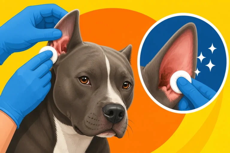
- Head and Face: Start with their head. Gently feel around their muzzle, under their eyes, and along their jawline. Check inside their ears (while also looking for signs of Pit Bull Ear Infections and What Drives Them) and around the base of their ears. Don't forget their neck, especially under the chin where lymph nodes are located.
- Neck and Shoulders: Move down their neck, feeling along the throat, and into their broad shoulders. Pay close attention to the armpits and chest area, as these are common spots for various types of lumps, including mast cell tumors.
- Torso and Back: Systematically move your hands across their rib cage, chest, and back. Use the pads of your fingers, applying gentle but firm pressure, moving in small circular motions. Feel along their spine and down both sides of their body.
- Belly and Groin: This area often has less hair, making lumps potentially easier to see, but harder to feel if they're small and soft. Gently part the fur and visually inspect, then carefully palpate their entire belly, including the groin area where lymph nodes are also present.
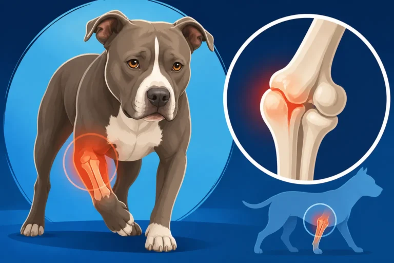
- Legs and Paws: Work your way down each leg, from shoulder/hip to paw. Feel all around the joints – elbows, knees, hocks – and along the muscles. Don't forget to check between their paw pads and their nail beds. These areas are prone to irritation and injury, but also potential lump development.
- Tail and Rear: Finally, check their tail from base to tip, and around their hindquarters, including under their tail and around the anal area.
The goal is to feel the underlying skin and tissue, not just stroke the fur. You're looking for anything that feels "new" or "different" from previous checks. Do this routine at least once a month. The more familiar you are with your Pit Bull's normal body, the quicker you'll spot something out of the ordinary. And remember, consistency is key. Make it a regular part of your grooming or petting routine.
What to Look For: Characteristics of Suspicious Lumps
So you've found a lump. Don't panic! Many lumps on dogs are benign. However, understanding what characteristics might raise a red flag for a mast cell tumor can help you communicate effectively with your vet and advocate for your Pit Bull.
Size, Texture, and Mobility
When you find a lump, take a moment to carefully assess its physical characteristics.
- Size: How big is it? Is it tiny, pea-sized, or larger? Crucially, has it changed size since you first noticed it? Mast cell tumors can sometimes grow rapidly, but they can also fluctuate in size, seemingly getting smaller only to re-enlarge later. This "waxing and waning" is a classic, albeit tricky, mast cell tumor characteristic.
- Texture: What does it feel like? Is it soft and squishy, like a fatty lipoma? Is it firm and rubbery? Or is it hard and unyielding, almost bony? Mast cell tumors can present with any of these textures, making them challenging to identify by feel alone. Some are firm and solid, others feel more gelatinous.
- Mobility: Can you move the lump freely under the skin, or does it seem attached to the underlying muscle or deeper tissues? Benign growths like lipomas (fatty tumors) are often very mobile. Mast cell tumors can be mobile, especially if they are superficial in the skin, but they can also be fixed and invasive, making them harder to shift around. If a lump feels rooted or attached, that's often a more concerning sign.
It's helpful to make a mental note, or even jot down, these characteristics. "Pea-sized, firm, freely movable lump on the left flank" gives your vet a much better starting point than "a lump on his side."
Location and Surrounding Skin Changes
The location of a lump can sometimes offer clues, though mast cell tumors truly can appear anywhere. They are most commonly found on the skin, but also frequently on the limbs, and less commonly on the trunk or head. However, don't let a "typical" location guide your concern too much, as atypical locations are always possible.
Beyond the lump itself, pay close attention to the skin directly over and around the growth. Are there any visible changes?
- Redness or Inflammation: Is the skin over the lump red, inflamed, or irritated? This can be a sign of infection, but it can also be a characteristic of a mast cell tumor due to the inflammatory chemicals they release.
- Ulceration or Crusting: Has the skin broken open? Is there a scab or crust? Ulcerated or weeping skin over a lump is always a red flag and warrants immediate veterinary attention, as it can indicate an aggressive tumor or a secondary infection.
- Hair Loss: Is there any hair loss directly over or around the lump?
- Swelling: Is the area around the lump swollen? This can also be a sign of inflammation or an aggressive growth.
A mast cell tumor might also cause what's known as Darier's sign, where rubbing the tumor causes it to redden and swell as it releases histamine. While not always present, if you observe this, it's a strong indicator.
Symptoms Beyond the Lump Itself
Sometimes, a mast cell tumor isn't just a local problem; it can cause systemic signs due to the chemicals (like histamine) it releases. These symptoms can be subtle or dramatic and are often linked to a higher-grade tumor or one that has spread. Keep an eye out for these potential red flags in your Pit Bull:
- Gastrointestinal Issues: Vomiting, diarrhea, or loss of appetite. These are classic signs of histamine release affecting the stomach and intestines. You might notice a sudden change in their eating habits, perhaps they're A Pit Bull That Refuses Food for a Day.
- Lethargy or Weakness: A general decrease in energy levels or unusual tiredness.
- Weight Loss: Unexplained weight loss, despite a normal appetite.
- Black, Tarry Stools: This is a serious sign, indicating digested blood in the stool, often due to stomach ulcers caused by histamine release from the tumor.
- Itching or Discomfort: The dog might scratch or lick the area excessively, even if the lump isn't visibly irritated. This could be local discomfort or a broader allergic-type reaction.
These broader symptoms, especially when coupled with the discovery of a new lump, should prompt an immediate veterinary visit. It's easy to dismiss some of these as minor ailments, but combined, they can paint a more serious picture.
Here's a quick comparison of common lumps you might find:
| Characteristic | Mast Cell Tumor (MCT) | Lipoma (Fatty Tumor) | Sebaceous Cyst | Wart/Papilloma |
|---|---|---|---|---|
| Appearance | Highly variable; can be red, inflamed, ulcerated | Soft, round, or oval, often under the skin | Small, firm or soft, skin-colored, may rupture | Cauliflower-like, rough, often pigmented |
| Texture | Soft, firm, rubbery, or hard | Soft, squishy, greasy feel | Firm or fluctuant (fluid-filled) | Firm, irregular surface |
| Mobility | Can be mobile or fixed to underlying tissue | Highly mobile, easily moved under the skin | Mobile, attached to skin | Fixed to skin |
| Size Fluctuation | Can "wax and wane" (change size) | Usually stable, slow growth | May increase/decrease if fluid builds/drains | Stable, or slow growth |
| Common Location | Skin (most common), limbs, trunk, head, internal | Trunk, armpits, groin, limbs | Head, neck, back, eyelids | Face, mouth, paws (especially in young dogs) |
| Associated Signs | Redness, swelling, itching, ulceration, GI upset | Generally none | May burst, discharge white/yellow material | Usually none, unless irritated |
| Urgency to Vet | High – needs prompt diagnosis | Monitor, vet for growth/concern | Vet if inflamed, growing, or bothering dog | Monitor, vet if growing rapidly or bleeding |
Next Steps After Finding a Lump on Your Pit Bull
Finding any new lump is a moment to take seriously. While we want to avoid unnecessary worry, being proactive is always the best approach when it comes to your Pit Bull's health.
When to See the Veterinarian
The short answer is: always. Any new lump or bump, especially one with concerning characteristics like those described above, warrants a veterinary examination. Even if you think it's "probably nothing," it's better to be safe than sorry. Mast cell tumors are notorious for their variable appearance, making it impossible to definitively diagnose them just by looking or feeling.
Don't wait if you notice:
- Rapid growth or any change in size (even getting smaller).
- Redness, swelling, ulceration, or bleeding.
- The lump appears to be bothering your Pit Bull (licking, scratching).
- Your Pit Bull is showing any systemic signs like lethargy, loss of appetite, or vomiting.
For Pit Bulls, given their breed predisposition, I recommend a vet visit for any new lump. Seriously. It might feel like you're overreacting, but catching a mast cell tumor early can be the difference between a simple removal and a much more complex, potentially life-threatening situation. Your vet would much rather tell you it's a benign cyst than hear you say you waited too long.
Diagnostic Tests for Lumps
Once you're at the vet, they'll perform a thorough physical exam, noting the lump's characteristics and location. The most common first step for diagnosing a lump is a Fine Needle Aspirate (FNA). This is a relatively quick, minimally invasive procedure where your vet uses a small needle (similar to one used for vaccinations) to collect a sample of cells from the lump. The cells are then spread on a slide and examined under a microscope.
An FNA can often, but not always, give a definitive diagnosis. Mast cell tumors have a distinctive appearance under the microscope, characterized by numerous mast cells with granular cytoplasm. However, if the FNA is inconclusive, or if the vet has a strong suspicion of a mast cell tumor despite a negative FNA, a biopsy may be recommended. A biopsy involves surgically removing a small piece of the lump, or even the entire lump, which is then sent to a pathology lab for more in-depth analysis. This provides not only a definitive diagnosis but also helps determine the tumor's grade (how aggressive it is) and margin status (if it was completely removed), which are crucial for prognosis and treatment planning.
Other tests might include blood work to check overall health, especially if systemic signs are present, and imaging (X-rays, ultrasound) if an internal tumor is suspected or if the vet wants to check for spread to lymph nodes or other organs.
Treatment Options and Prognosis
If a mast cell tumor is diagnosed, the treatment plan will depend heavily on the tumor's grade, location, and whether it has spread. For most skin-based mast cell tumors, surgical removal is the primary and often curative treatment. The goal is to remove the tumor with "wide margins," meaning a significant amount of healthy tissue around and beneath the tumor, to ensure all cancerous cells are gone. This can sometimes result in a larger incision than anticipated, especially if the tumor is on a limb or an area with less skin elasticity.
For higher-grade tumors, or those that have spread, additional treatments may be recommended, such as:
- Radiation Therapy: Used to target any remaining cancer cells after surgery, or for tumors that cannot be completely removed surgically.
- Chemotherapy: Often used for high-grade tumors, those that have spread, or for cases where surgery alone isn't sufficient. There are various chemotherapy protocols, tailored to the individual dog.
- Targeted Therapies: Newer drugs like Palladia or Kinavet are oral medications that target specific pathways involved in cancer growth, offering another option for certain types of mast cell tumors.
The prognosis for mast cell tumors varies widely. Low-grade tumors, completely removed with wide margins, often have an excellent prognosis. High-grade tumors, or those that have spread, carry a more guarded prognosis, but advancements in veterinary oncology mean that even these cases can often be managed effectively, extending quality of life. Regular follow-up appointments and ongoing monitoring will be crucial after any treatment.
It’s important to remember that these treatments can be costly. This is where having pet insurance can truly be a lifesaver. Providers like Healthy Paws can help alleviate the financial burden of diagnostics, surgery, and ongoing treatments for conditions like mast cell tumors, allowing you to focus on your Pit Bull's recovery without added stress about the vet bills. While it can be a significant upfront investment, the peace of mind knowing you can make the best medical decisions for your dog without financial constraints is invaluable. If you're managing a chronic health issue like Pit Bull Skin Allergies and the Atopy Pattern, you already know how quickly vet bills can add up, and cancer treatments are often far more extensive.
Regular at-home checks combined with swift veterinary action at the first sign of trouble are your Pit Bull's best defense against mast cell tumors. You know your dog best, and your vigilance can make all the difference.
Disclaimer
This article is intended for informational purposes only and does not constitute veterinary advice. Always consult with a qualified veterinarian for any health concerns or before making any decisions about your pet's medical care.